With MRI Wellness, get the full picture of your wellbeing.

In doctors’ offices, we’re typically met with treatment methods – not prevention strategies. MRI Wellness has the solution. Through our MRI technology, we can catch conditions before they become a greater concern.
With a proactive approach to your health, our innovative MRI system delivers a comprehensive report, assessing your risk of disease, detecting early signs of cancer, and allowing you to dive deeper into the state of your wellbeing.
Our full body scan can help identify causes for unexplained symptoms, monitor genetic conditions or family history health risks, detect cancer and other illnesses, and maintain a proactive mindset for your health.
We check for hundreds of conditions and continue to make new discoveries every day.
An Unmatched MRI Experience
Our MRI experience is engineered for accuracy, speed, safety, and most of all comfortability. Above all, our machines are configured for diagnostic quality, providing comprehensive whole body imaging.

All eyes on your health
Each report is carefully deciphered by a member of our team, who will explain your results with context for both you and your doctor.
Get the full scope
Our team specializes in whole-body imaging rather than focusing on specific organs.
Sit back and relax
We’ve designed our MRI experience to greatly reduce anxiety associated with this type of appointment.
“Our mission is to make MRI scanning easily accessible for the early detection of disease, early cancers, and silent killers such as aneurisms.”

Pick out the right scan for you, then choose a day and time that works best for your schedule. We offer three choices: a whole-body scan, a brain and spine scan, or a torso scan.

On the day of your scan, we’ll check you in, confirm your physician’s information to send your report, give you something comfortable to wear, and get started on your full-body scan. You’ll be able to relax in our comfortable imaging space while our innovative MRI technology works its magic. In approximately an hour, you’ll be finished with your scan.

Once your results are in, one of our specialists will walk you through our findings. You can also share the information we gather with other members of your healthcare team, like your primary care physician or any specialists.
Experience detailed imaging of the entire body, from head to toe. This advanced scan covers major organs and systems, including the brain, spine, heart, lungs, and more!
This specialized scan provides detailed images of the spinal cord, vertebrae, discs, and surrounding soft tissues. It’s commonly used to diagnose and assess conditions like herniated discs, spinal stenosis, tumors, infections, and more.
Capture detailed images of the abdomen and pelvis and surrounding tissues.
This scan targets the regions most vulnerable to environmental exposure and inhalation, capturing high-resolution images of the brain, abdomen, and pelvis.

“You can’t put a price tag on the peace of mind that comes from truly knowing where your health stands.”
MRI Wellness has contributed to my sense of well-being a great deal. It has given me peace of mind and helped me feel empowered about my health.
“I had been suffering from constant headaches and migraines for months. I tried everything and still couldn’t get answers so I decided to go to MRI Wellness to figure out what everyone else was missing. The scan was comfortable, the team was professional and kind, and the results were explained in a way I could actually understand. It felt validating to finally get a clear picture of what was happening with my health.”
As a college student, it’s easy to assume you’re healthy—but actually seeing what’s going on inside your body changes your perspective. MRI Wellness gives me confidence that I’m on solid ground.
“I lived with severe back pain for years and had a procedure to straighten my spine, but I was still in pain afterward. I got a full-body MRI at MRI Wellness to figure out what was wrong. The staff made me feel so comfortable, and seeing my results gave me a sense of control and peace of mind again. I’m so grateful for this experience.”
“If you don’t know how your body is functioning on the inside, you don’t really know that you’re healthy—and that knowledge changes everything..”
“Knowing what’s happening inside my body gave me peace of mind—and the confidence to take better care of my health now, not later.”
As a mother, staying healthy for my family is my top priority. This became even more important to me when I considered my family history, as my grandmother battled ovarian cancer. Getting a full-body MRI was my way of ensuring I’m here for my family.
Absolutely! Health experts recommend whole-body MRI scans for people at high risk or those with a family history of certain health conditions. Many people simply want peace of mind regarding their health, which is understandable. While this is a new advancement in healthcare technology and research is ongoing, a full-body MRI is an excellent first step in assessing your overall health. It allows you to seek specialized medical assistance with a certified physician to properly address any health concerns detected.
Our full-body MRI scans are easily performed, painless procedures that deliver timely results to help you better understand your current health status. The scan provides information on every system and organ in the body and identifies any abnormal findings. You will receive a detailed report for your physician to review.
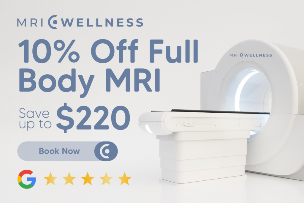
You can make an appointment online at MRI-WELLNESS.COM or by calling 1-833-WELLMRI.
MRI Wellness scans do not expose you to any harmful ionizing radiation like that used in X-rays, PET scans, or CT scans. In addition, MRI Wellness scans do not use contrast dyes.
MRI uses radio waves and a strong magnetic field to produce images of soft tissue and bone. An MRI is better at differentiating between normal and abnormal soft tissue, spotting injuries and musculoskeletal issues. CT scans use X-rays to produce images that help to show the edges of structures. They are ideal in emergency situations.
No. We can arrange a referral from an independent medical practitioner at no additional fee. In the event there is a serious medical finding, a medical practitioner can additionally help you understand and explain the findings in your MRI Wellness report.
Depending on the type of scan you choose, time in the MRI machine will vary from 30 to 60 minutes. If movement is detected, the scan may take slightly longer.
There are no evidence-based guidelines specifically recommending a total body MRI screening in the general population. This decision should be based on patient concerns, genetics, family history, and input from your primary healthcare provider. Statistics show that cancer risk, as well as any disease risk, increases with age.
Yes. At MRI Wellness, we only screen patients 18 years of age or older.
Our calm, comfortable clinic is designed for you to sit back, relax, and let us dedicate time to your wellness. Your health deserves the best.
2683 Elms Plantation Blvd North Charleston, SC 29406
Monday: 8:00 a.m. – 4:30 p.m.
Tuesday: 8:00 a.m. – 4:30 p.m.
Wednesday: 8:00 a.m. – 4:30 p.m.
Thursday: 8:00 a.m. – 4:30 p.m.
Friday: 8:00 a.m. – 3:00 p.m.
Saturday: Upon Request

Email:
info@MRI-Wellness
Tel:
1-833-WellMRI (1-833-935-5674)
Take the first step toward gaining a full picture of your health with MRI Wellness.